
Skin Anatomy & Physiology

The skin is the largest organ of the body. It is an elastic protective covering, which is thinnest on the lips and eyelids, and thickest on the palms of the hands and soles of the feet.

Structure of the skin: three major divisions

  • Epidermis – the outermost layer of the skin
  • Dermis – the inner corium made up of connective tissue, capillaries, nerve endings, and ECM
  • Subcutaneous – the deep layer of adipose (fatty) tissue
Skin anatomy illustration

The Structure of Human Skin

Epidermis

This is the outermost layer of the skin, which forms the protective covering of the body. Keratinocytes, or squamous cells, are produced in the basal layer (stratum germinativum) of the skin. These cells then migrate up through the epidermis until they complete their life cycle in the dead stratum corneum. The epidermis contains the following layers:

Basal layer (stratum basale) – consists of keratinocytes that undergo cell division and are responsible for the growth of the epidermis. This layer also contains melanocytes that protect the cells from the UV rays of the sun.

Spinous layer (stratum spinosum) – consists of multiple layers of square-shaped cells. The desmosomes that hold the cells together have a spiny or prickle-like appearance when viewed under a microscope. As cells move upward during natural cell turnover, they become more flat. This layer also contains Langerhans cells that assist in the skin’s immune function.

Granular layer (stratum granulosum) – consists of dying cells that contain distinct granules.

Clear layer (stratum lucidum) – is a barrier composed of transparent cells through which light can pass. These cells are only present in thick skin, such as the palms of the hands and soles of the feet.

Cornified layer (stratum corneum) – is composed of tightly packed dead cells, which are constantly being shed and replaced. This layer contains the skin’s natural moisturizing factor (NMF) and is responsible for maintaining hydration of the stratum corneum. NMF exists inside the corneocytes (dead keratinocytes).

The NMF includes a combination of:

  • amino acids
  • sodium PCA
  • lactate
  • urea
  • ammonia
  • uric acid
  • glucosamine
  • creatinine
  • organic acids
  • citrate
  • sodium potassium
  • calcium
  • magnesium
  • phosphate
  • chlorine
  • sugar
  • peptides
  • other unidentified substances

A decrease in the NMF results in an increase in transepidermal water loss (TEWL), causing dryness and inflammation. The protein keratin and the lipid bilayer around each cell also assist in hydration by acting as occlusives, waterproofing the skin and preventing water evaporation. A small amount of TEWL is constantly taking place within the cornified layer and is critical to skin health; however, TEWL increases when the epidermis is compromised and may result in sensitivity.

Dermis

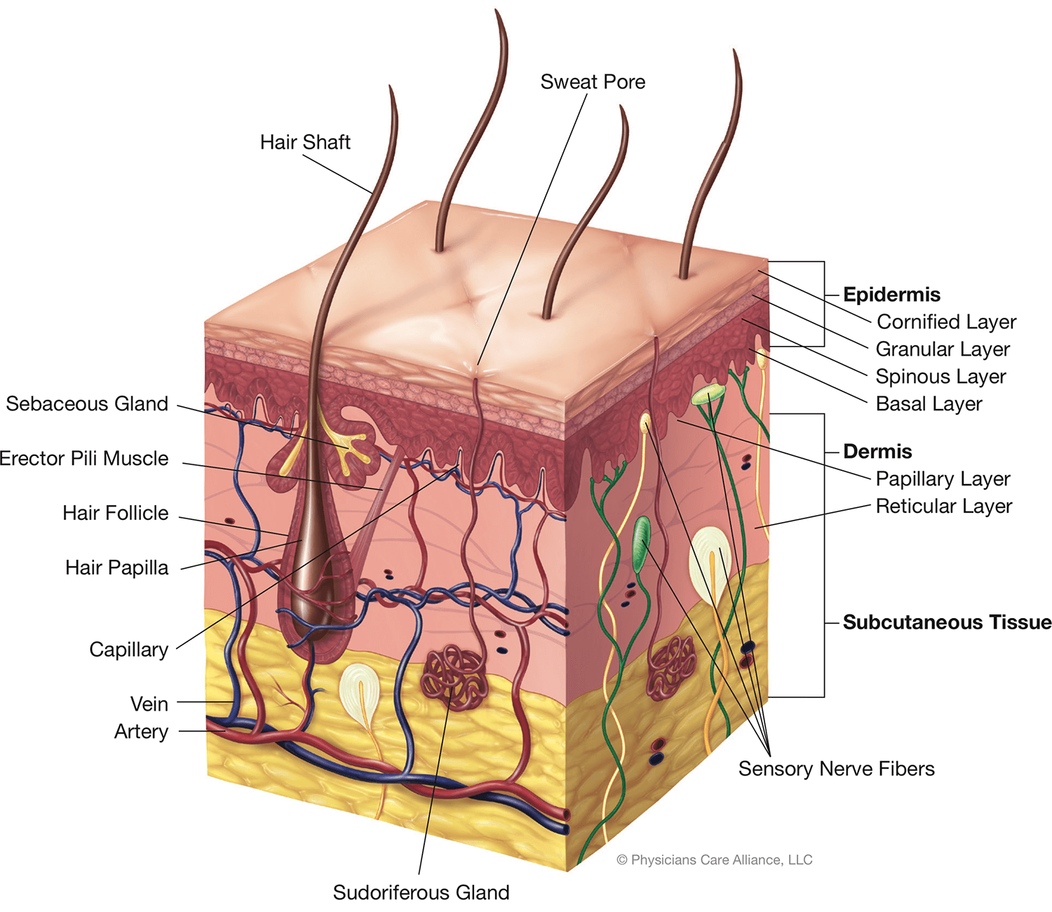
The dermis is the layer lying underneath the epidermis, consisting of highly sensitive and vascular connective tissue, collagen, elastin, and reticular fibers. It consists of two layers: the papillary layer and the reticular layer.

Papillary layer – is composed of small conical elevations called “papillae,” which push up into the epidermis and contain blood vessels and nerve fibers called tactile corpuscles. The epidermis receives its nourishment through this blood supply. Through these nerve fibers, we gain our sense of touch.

Reticular layer – contains the extracellular matrix (ECM), blood vessels, lymph nodes, nerves, sebaceous and sudoriferous glands, and fibrous and elastic tissue.

Extracellular matrix (ECM) – is a complex group of biomolecules designed to support and protect the cells. The ECM consists of structural proteins, such as collagen; adhesive proteins, such as fibronectin; glycosaminoglycans (GAG), such as hyaluronic acid; and elastin. A healthy ECM provides the skin with a firm and youthful appearance; however, environmentally-induced enzymes in the skin known as matrix metalloproteinase (MMP) break down and recycle proteins and can prematurely degrade this structure.

Matrix metalloproteinase (MMP) – are enzymes such as collagenase and elastase, and are responsible for the breakdown and recycling of proteins like collagen and elastin. MMP are beneficial to a certain extent and fight environmental factors such as pollution and other free radicals. When too many of these enzymes are present, the result is an unwanted breakdown of healthy proteins and extrinsic aging.

Matrix metalloproteinase inhibitors (MMPi) – are ingredients that maintain the correct balance within the extracellular matrix by inhibiting the activity of the enzymes responsible for the breakdown of its structure (MMP). Examples of common MMPi are antioxidants such as resveratrol, epigallocatechin gallate (EGCG), and vitamins C and E.

Sebaceous glands – are small oil-producing glands that consist of little sacs whose ducts open into the hair follicles. They secrete sebum, which lubricates and prevents moisture loss from the skin. These glands are found in all parts of the body with the exception of the palms of the hands and soles of the feet. These glands respond to androgen hormones (male hormones) and increase the production of sebum during adolescence.

Sudoriferous glands – are tubular glands that are abundant in most parts of the body, but are more predominant on the palms, soles, forehead, and underarms. The sudoriferous glands regulate body temperature and help eliminate waste products from the body. There are two types of sudoriferous glands found in the skin: eccrine and apocrine glands.

  • Eccrine glands – secrete a watery sweat that is a mixture of 99% water and 1% salts and fats. They are not associated with the hair follicle and respond to temperature to keep the body cool.
  • Apocrine glands – become active at puberty. These glands are larger and deeper than eccrine glands and are associated with the hair follicle. Apocrine glands produce thicker secretions than eccrine glands and contain pheromones, which emit odor.

Subcutaneous Layer

This is the fatty layer lying directly below the dermis, which is composed of fat cells, blood, nerves, and lymph supply. It gives smoothness and contours to the body, contains fat that is utilized by the body for energy, and acts as a protective cushion for the outer skin.

Functions of the Skin

The following are the six primary functions of the skin:

  1. Heat regulation
    The skin regulates the body temperature by sweating. Sweat is produced by the sudoriferous glands. The evaporation of the moisture enables the body to cool itself. Surface capillaries dilate, resulting in flushing and the release of heat.
  2. Absorption
    The epidermis contains an acid mantle layer, which limits the amount of substances entering through the skin. Cosmeceutical treatment products must be formulated at an appropriate pH with chirally correct ingredients for optimal penetration and absorption.
  3. Secretion
    The sebaceous glands excrete sebum to lubricate and maintain the health of the skin.
  4. Protection
    The fat cells in the adipose layer provide insulation and protection against trauma to the internal organs. The skin also protects itself from the harmful effects of UV rays and light, and acts as a barrier against the invasion of bacteria.
  5. Excretion
    Perspiration is the process by which the sudoriferous (sweat) glands excrete waste materials, water, and salt.
  6. Sensation
    Nerve endings in the skin allow us to feel heat, cold, touch, pleasure, pressure, itch, and pain.